Spatial transcriptomics was named Method of the Year in 2020 by Nature, recognizing its fundamental contribution to understanding the intricate genetic relationships that govern our health. Today, spatial transcriptomics continues to improve our understanding of various diseases. In particular, cancer research has benefitted greatly from the use of spatial transcriptomics. It has enabled researchers to understand the underlying mechanisms of cancer development and how specific treatments affect gene expression in tumor cells.
A core part of this method is fluorescence scanning, as it enables the visualization of gene expression patterns in a tissue sample. However, fluorescence microscopy is currently limited by the optics, throughput, and complexity of microscope systems. To keep pace with the current and future needs of researchers, microscopy core facilities worldwide are looking to increase automation, throughput, and device simplicity, with the goal of making analysis more efficient without compromising on image quality.
We recently interviewed Dr. Alvaro Crevenna, the head of microscopy at EMBL Rome, about the development of new methods in spatial transcriptomics. He discussed how the SLIDEVIEW™ VS200 research slide scanner from Evident has been essential for technology development and increasing throughput as well as extending the current capabilities of fluorescence
scanning.
Processing High Volumes of Tissue Samples
“The bread and butter of the imaging needs here is tissue imaging” — Dr. Alvaro Crevenna, the head of microscopy at EMBL Rome
EMBL Rome's head of microscopy, Alvaro Crevenna, leads a facility that helps people image their samples. The facility focuses on two research topics: epigenetics and neuroscience, both of which involve working with mice. Tissue imaging is a crucial need for the facility, with fixed tissue accounting for 99% of samples. When Alvaro arrived in 2019, the facility had three instruments, but none of them were tailored to tissue imaging. This meant that researchers were spending an inordinate amount of time processing samples.
This was especially true for neurobiology researchers. In neuroscience research, visualizing neuronal activity is crucial to understanding the workings of the brain. Researchers often use mini scopes or two photon scopes to monitor and record neuronal activity during a set of behaviors and tasks. After the experiment, the first step is to section the brain and check whether the virus was injected correctly. However, it was taking a long time for researchers to manually locate the areas that needed to be imaged and select regions of interest (ROI) for subsequent high-resolution scans.
Therefore, researchers needed a more efficient and accurate way of identifying ROIs, and a system that was specifically for processing high volumes of tissue samples.
The Need to Automate Tissue Imaging
When looking to find the best slide imaging system for the microscopy facility, Alvaro considered and trialed several machines, including the VS200 research slide scanner from Evident.
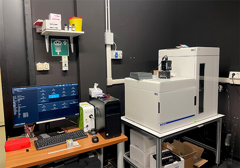
The VS200 research slide scanner at EMBL Rome.
The VS200 scanner was chosen due to its remarkable flexibility, which enables simultaneous overview and fluorescence scanning. In addition, the software's AI-guided tissue detection algorithm simplified tissue scanning and ROI detection, reducing time and effort. These were both key factors for the EMBL microscopy facility, as Alvaro noted:
“One of the really beautiful things about the VS200 research slide scanner from Evident is that you can perform the overview image with fluorescence.” — Dr. Alvaro Crevenna, the head of microscopy at EMBL Rome
In addition to automated image processing, the VS200 scanner offers a higher capacity and throughput for processing slides than many comparable models. Alvaro found that researchers can now accomplish 10x more tasks by running processes overnight, eliminating several hours of manual slide processing during the working day. The VS200 scanner also demonstrates exceptional image quality and optics and is incredibly easy to use. Alvaro states, “Normally in a 40-minute training session, people are up and running on their own.” This is an important point as it means that users of all abilities can quickly become confident with the system, both increasing accessibility of the equipment and reducing the training burden on the imaging facility.
These features were critical to Alvaro's work at EMBL, where they are developing innovative technologies, including the implementation of in situ sequencing for spatial transcriptomics. The VS200 scanner will be a vital tool for EMBL's future research, helping them achieve their goals with ease and efficiency.
Extending the Boundaries of Spatial Transcriptomics
Spatial transcriptomics is a powerful technology that enables the visualization of gene expression patterns in situ, providing insights into cellular diversity and organization in tissues. However, the number of channels that may be observed often restricts the technique's ability to investigate a greater number of genes in a single tissue segment.
Alvaro’s team at EMBL Rome recognizes the need to improve the capabilities of spatial transcriptomics, particularly in neuroscience and epigenetics research. To achieve this, the facility aims to extend the number of wavelengths that can be visualized and develop automated analysis pipelines for efficient data processing.
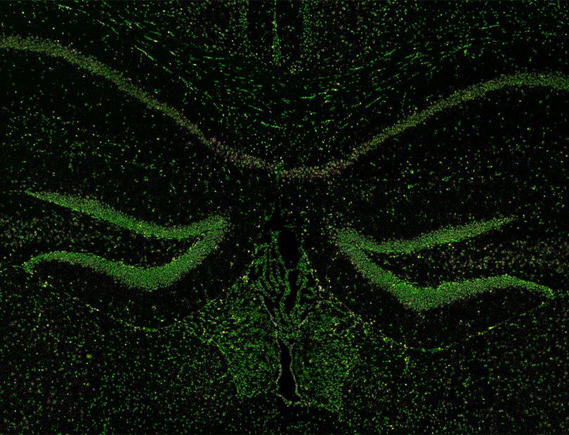
Using 3 distinct fluorescence channels (DAPI, Cy3, and Cy5), spatial transcriptomics reveals the presence of RNA molecules within a coronal mouse brain section.
Traditionally, microscopes are limited to visualizing about five different wavelengths. With the capacity for multiplexing and combined observational methods, the VS200 scanner is a prime instrument to extend the number of wavelengths that can be reliably visualized. This lets researchers observe the activity of more genes in the tissue, reducing the number of imaging cycles while improving the quality and accuracy of their results.
Through collaboration with Evident, Alvaro plans to explore the potential of multiplexing to further extend the number of channels. Currently, they have designed a system that can visualize up to 11 channels, providing further detailed insights into gene expression patterns in tissues. This number is likely to increase as the collaboration continues.
To make this research accessible to others, the facility is also developing automated analysis pipelines. These pipelines will streamline data processing, enabling researchers to analyze their results quickly and efficiently. Additionally, Alvaro is exploring the implementation of basic computational super-resolution methods to further enhance the quality of data:
“As a facility, we believe that our work should be done in a way that others can adopt it. We are already discussing the implementation of our new algorithm for spectral unmixing with Evident, with the goal of making it available to everyone who conducts this type of research.”
By extending the boundaries of spatial transcriptomics through multiplexing and automated analysis pipelines, EMBL Rome is advancing the field of neuroscience and epigenetics. They are also providing researchers with the tools they need to gain deeper insights into gene expression patterns in tissues.
Future Visions
EMBL Rome has a clear vision for the future use of the VS200 scanner, which will play a pivotal role in the development of the spatial transcriptomic service platform. Rather than automating a single sample, EMBL Rome plans to parallelize the sample preparation process, which will enable a higher throughput of slides on the VS200 scanner. This will enable EMBL Rome to increase the efficiency of their experiments while maintaining high levels of accuracy and precision. By using the VS200 scanner
in this manner, EMBL Rome hopes to achieve their goal of creating a platform that can provide high-quality spatial transcriptomic data to researchers all over the world.
Related Content
Identifying Cirrhosis in Liver Tissue Using a Research Slide Scanner
From Multimode Imaging to Multiplexing: Your Essential Guide to Extracting Rich Data